41 heart structure and labels
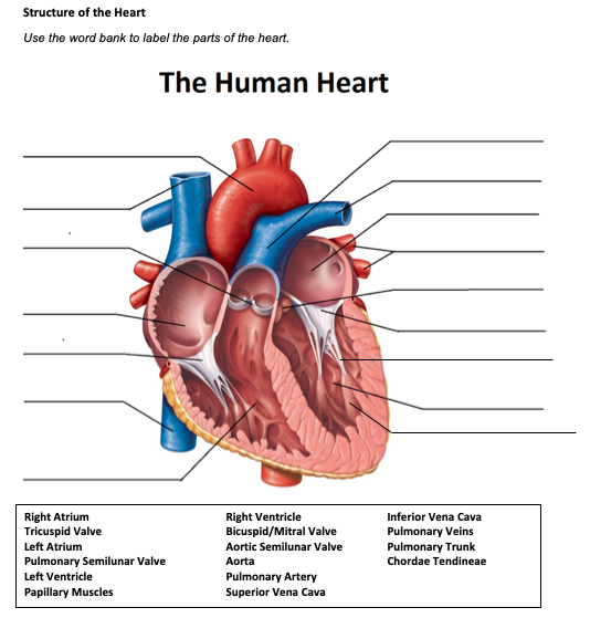
How the Heart Works - The Heart | NHLBI, NIH - National Institutes of ... The Heart. The heart is an organ about the size of your fist that pumps blood through your body. It is made up of multiple layers of tissue. Your heart is at the center of your circulatory system. This system is a network of blood vessels, such as arteries, veins, and capillaries, that carries blood to and from all areas of your body. Reinforcement: Anatomy of the Human Heart - The Biology Corner Reinforcement: Anatomy of the Human Heart. I created this worksheet for my anatomy students to review the anatomy of the heart. Students focus on vocabulary that relates to anatomical features, such as the mitral valve, aorta, and heart chambers. At the end, students label a line drawing of the heart. I included a word bank at the top, but you ...
Free Heart Worksheets for Human Anatomy Lessons - Homeschool Giveaways Print out sheet of the human heart with labels - This fun heart worksheet shows kids the different parts of the heart. They'll learn about the left ventricle, the left atrium, the tricuspid valve, and more. Human Heart Clipart - There is a coloring page, heart labeling worksheet and heart anatomy chart.
Heart structure and labels
How the Heart Works: Diagram, Anatomy, Blood Flow - MedicineNet The heart is about the size of a closed fist, weighs about 10.5 ounces, and is somewhat cone-shaped. It is covered by a sack termed the pericardium or pericardial sack. The normal heart anatomy consists of a four-chambered, hollow organ. It is divided into the left and right sides by a muscular wall called the septum. Lab 2: Anatomy of the Heart - Anatomy & Physiology: BIO 161 / 162 ... Lab 2: Anatomy of the Heart; Search this Guide Search. Anatomy & Physiology: BIO 161 / 162. AP BIO 161 / 162; AP 1: BIO161 Toggle Dropdown. Chapter 1: An Introduction to the Human Body ; Chapter 4: The Tissue Level of Organization ; Chapter 5: The Integumentary System ; 20 Free Printable Heart Templates, Patterns & Stencils Here's how to make a paper heart chain with our heart template. All you need is a printer and some scissors. Step 1: Download & print the template Download and print our paper heart chain template. The template can make two heart chains per page, and the hearts will be approximately 2 inches in height. Paper Heart Chain Template
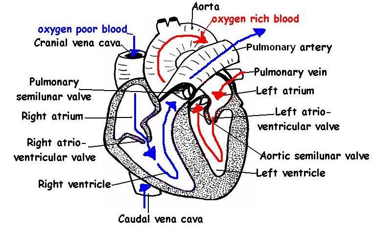
Heart structure and labels. Blank Heart Diagram To Label - The Heart Simple Teaching Resources Label heart interior anatomy diagram printout. These human heart diagrams are . In this interactive, you can label parts of the human heart. Furthermore, a blank human heart diagram has been provided, allowing you to fill in the blanks for an incoming test or quiz. Download this free simple heart diagram to label and help your class learn all ... Parts of the Heart: How does blood flow through it? - Study.com The four parts of the heart are the right atrium, right ventricle, left atrium, and left ventricle. The atrium receives blood from the body, the right ventricle pumps blood to the lungs, the left ... 40.9: Mammalian Heart and Blood Vessels - Biology LibreTexts Structure of the Heart. The heart is a complex muscle that pumps blood through the three divisions of the circulatory system: the coronary (vessels that serve the heart), pulmonary (heart and lungs), and systemic (systems of the body). Coronary circulation intrinsic to the heart takes blood directly from the main artery (aorta) coming from the ... Chapter 27 - Heart Anatomy - BIO 140 - Human Biology I - Textbook ... The human heart consists of four chambers: The left side and the right side each have one atrium and one ventricle. Each of the upper chambers, the right atrium (plural = atria) and the left atrium, acts as a receiving chamber and contracts to push blood into the lower chambers, the right ventricle and the left ventricle.
Physiology, Cardiac - StatPearls - NCBI Bookshelf Many of these defects arise from birth and have significant pathology that alters the structure of the heart. The most common cardiac defect is a ventricular septal defect (VSD) that is essentially an opening in the septum between the ventricles. From birth, this defect causes a shunt from the left side of the heart (higher pressure) to the ... Diagrams, quizzes and worksheets of the heart | Kenhub Labeled heart diagrams Take a look at our labeled heart diagrams (see below) to get an overview of all of the parts of the heart. Once you're feeling confident, you can test yourself using the unlabeled diagrams of the parts of the heart below. Labeled heart diagram showing the heart from anterior Unlabeled heart diagrams (free download!) Cardiovascular System 1, Heart, Structure and Function Cardiovascular System 1, Heart, Structure and Function Health Care November 07, 2021 ... Human heart: Anatomy, function & facts | Live Science The human heart has four chambers: two upper chambers (the atria) and two lower ones (the ventricles), according to the National Institutes of Health. The right atrium and right ventricle together...
Body Cavities and Membranes: Labeled Diagram, Definitions - EZmed The cranial cavity is the superior portion of the dorsal cavity, as we can see highlighted in red and labeled by the star below. The cranial cavity is enclosed by the cranium or skull, and it houses the brain . The cranial cavity is filled with fluid called cerebrospinal fluid that helps protect and cushion the brain. The Human Heart - Anatomy & Passage Of Blood - TeachPE.com The heart pumps continuously, without resting and without becoming fatigued. Its function is to pump blood to the lungs and around the body. The heart is one of the key organs in the Circulatory System. Anatomy of the heart. The heart consists of four chambers and is divided into left and right by a wall of muscle called the septum. Heart: Anatomy | Concise Medical Knowledge - Lecturio Heart: Anatomy. The heart is a 4-chambered muscular pump made primarily of cardiac muscle tissue. The heart is divided into 4 chambers: 2 upper chambers for receiving blood from the great vessels, known as the right and left atria, and 2 stronger lower chambers, known as the right and left ventricles, which pump blood throughout the body. Correctly Label The Following Internal Anatomy Of The Heart The aorta, or aortic arch, is the outermost layer of the heart. The left ventricle is covered with the ventricular aorta, and the pulmonary veins are located inside the aorta. The two atria, the left and right aorta, and the right aortic arch are all external organs. These organs carry oxygen-rich blood to the body.
The Heart - Labelled diagram
Mnemonics for Heart Anatomy and Physiology (Video) - Mometrix A heart block is an abnormal heart rhythm known as an arrhythmia and can occur anywhere in the specialized conduction system of the heart. The electrical signals telling the heart to contract are partially or totally blocked between the atria and ventricles. Therefore, it is called an atrioventricular, or AV block.
31 Human Heart To Label - Labels Design Ideas 2020
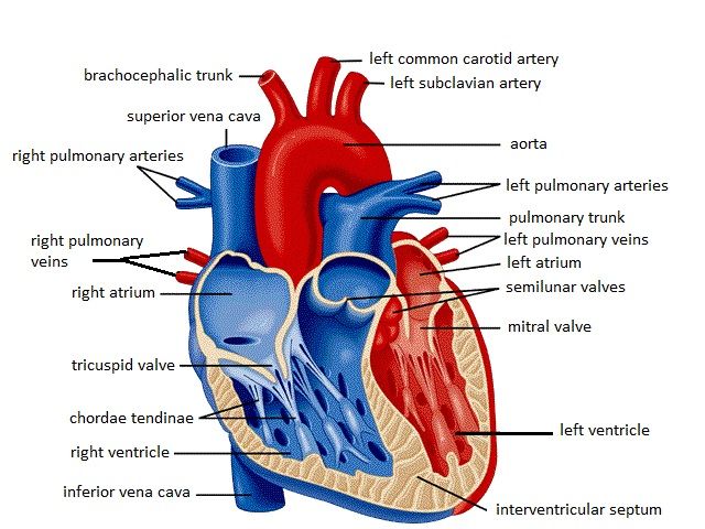
Heart anatomy: Structure, valves, coronary vessels | Kenhub Inside, the heart is divided into four heart chambers: two atria (right and left) and two ventricles (right and left).

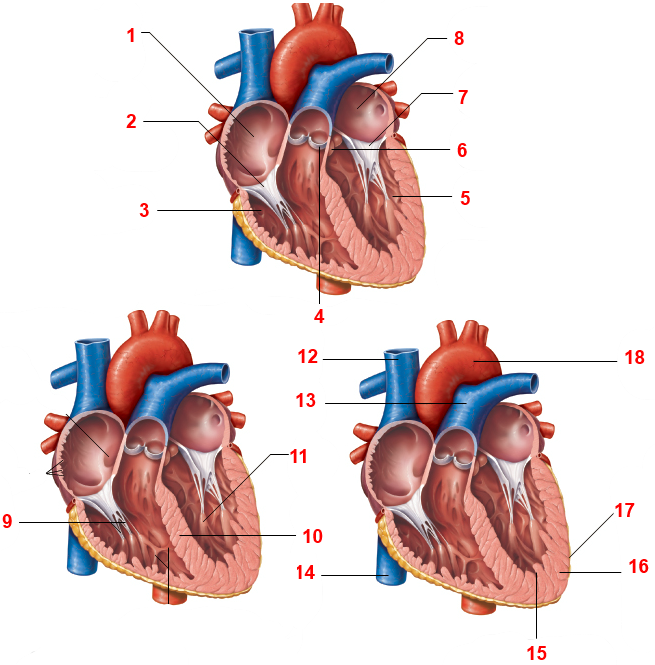
Fig 2 Gross Anatomy of the Heart (c)
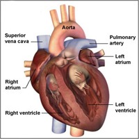
Know the Structures and Functions about Your Heart The heart has four chambers: Right atrium Left atrium Right ventricle Left ventricle Atria are smaller than ventricles and have thin, less muscular walls. They are the receiving chambers of the blood, which is delivered to the heart by the large veins. Ventricles are the larger, more muscular pumping chambers that push blood out to the circulation.

The brain - structure and function - Cancer Information - Macmillan Cancer Support
Histology, Heart - StatPearls - NCBI Bookshelf The heart is a four-chambered organ responsible for pumping throughout the body. It receives deoxygenated blood from the body, sends it to the lung, receives oxygenated blood from the lungs, and then distributes the oxygenated blood throughout the body. At the histological level, the cellular features of the heart play a vital role in the normal function and adaptations of the heart.
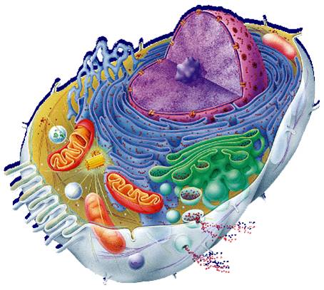
Free Animal Cell Unlabeled, Download Free Clip Art, Free Clip Art on Clipart Library
Heart Labeling Quiz: How Much You Know About Heart Labeling? Here is a Heart labeling quiz for you. The human heart is a vital organ for every human. The more healthy your heart is, the longer the chances you have of surviving, so you better take care of it. Take the following quiz to know how much you know about your heart. Questions and Answers 1. What is #1? 2. What is #2? 3. What is #3? 4. What is #4?
Heart Functions, Heart Diseases and Structure with Diagram
Identify Various Parts Of A Human Heart: Trivia Quiz It is in charge of keeping the processes within the body moving by facilitating the transfer of blood throughout the body. The quiz below is to test out interesting facts you may know about the heart. Give it a try and good luck. Questions and Answers 1. What is F? 2. What is K? 3. What is C? 4. What is D? 5. What is J? 6. What is E? 7. What is i?
labelled diagram of heart a level - Clip Art Library
EKG Interpretation & Heart Arrhythmias Cheat Sheet - Nurseslabs Use this EKG interpretation cheat sheet that summarizes all heart arrhythmias in an easy-to-understand fashion. One of the most useful and commonly used diagnostic tools is electrocardiography (EKG) which measures the heart's electrical activity as waveforms. An EKG uses electrodes attached to the skin to detect electric current moving ...
heart labelled diagram | Diabetes Inc.
heart | Structure, Function, Diagram, Anatomy, & Facts A thin layer of tissue, the pericardium, covers the outside, and another layer, the endocardium, lines the inside. The heart cavity is divided down the middle into a right and a left heart, which in turn are subdivided into two chambers. The upper chamber is called an atrium (or auricle), and the lower chamber is called a ventricle.

Lable Heart : New users enjoy 60% off. - Magic Pau
Heart: illustrated anatomy - e-Anatomy - IMAIOS Anatomical structures were labelled according to the actual Terminologia Anatomica. Anatomy of the human heart and coronaries: how to visualize anatomic structures This tool provides access to several medical illustrations, allowing the user to interactively discover heart anatomy.
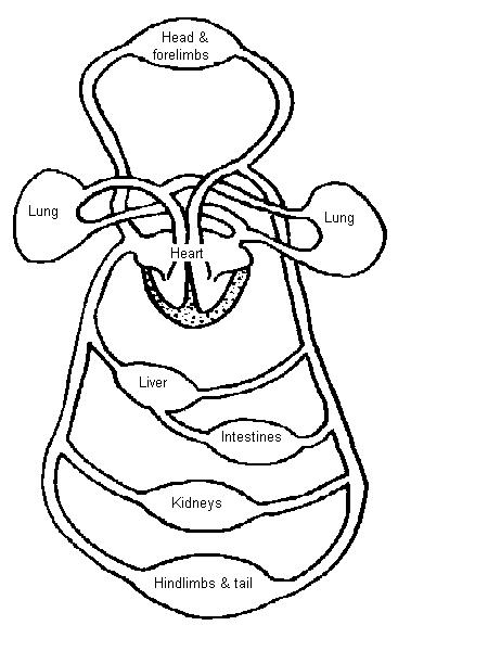
The Anatomy and Physiology of Animals/Circulatory System Worksheet - WikiEducator
Structure and Function of the Heart - News-Medical.net Structure of the heart The heart wall is composed of three layers, including the outer epicardium (thin layer), middle myocardium (thick layer), and innermost endocardium (thin layer). The...

Katelyn's Anatomy. .: HEART DISSECTIONS
Diagram of Human Heart and Blood Circulation in It A heart diagram labeled will provide plenty of information about the structure of your heart, including the wall of your heart. The wall of the heart has three different layers, such as the Myocardium, the Epicardium, and the Endocardium. Here's more about these three layers. Epicardium

The chest x-ray in cardiovascular disease - wikidoc
Heart - Wikipedia The heart has four chambers, two upper atria, the receiving chambers, and two lower ventricles, the discharging chambers. The atria open into the ventricles via the atrioventricular valves, present in the atrioventricular septum. This distinction is visible also on the surface of the heart as the coronary sulcus.[18]
The heart - Teaching resources
20 Free Printable Heart Templates, Patterns & Stencils Here's how to make a paper heart chain with our heart template. All you need is a printer and some scissors. Step 1: Download & print the template Download and print our paper heart chain template. The template can make two heart chains per page, and the hearts will be approximately 2 inches in height. Paper Heart Chain Template

QuickStudy | Nervous System Laminated Study Guide | Nervous system, Heart attack symptoms, Anatomy
Lab 2: Anatomy of the Heart - Anatomy & Physiology: BIO 161 / 162 ... Lab 2: Anatomy of the Heart; Search this Guide Search. Anatomy & Physiology: BIO 161 / 162. AP BIO 161 / 162; AP 1: BIO161 Toggle Dropdown. Chapter 1: An Introduction to the Human Body ; Chapter 4: The Tissue Level of Organization ; Chapter 5: The Integumentary System ;

Leadership: Group Work Can Be As Successful As You Want It To Be
How the Heart Works: Diagram, Anatomy, Blood Flow - MedicineNet The heart is about the size of a closed fist, weighs about 10.5 ounces, and is somewhat cone-shaped. It is covered by a sack termed the pericardium or pericardial sack. The normal heart anatomy consists of a four-chambered, hollow organ. It is divided into the left and right sides by a muscular wall called the septum.
Labeling the Heart (Part Three) Quiz - By dilatory

13 Best Images of Hip Anatomy Of The Worksheet - Sunflower Anatomy Diagram, Anatomy Knee ...
Post a Comment for "41 heart structure and labels"