40 human eye diagram without labels
Human Body Diagram - Bodytomy The head, neck, torso, a pair of arms and legs, respectively constitute the external view of the body, often described as the superficial, first-layer of the human body. However, internally, the structure is far complex and intricate. Know that there are 11 organ systems of the body: Circulatory System, Respiratory System, Immune System ... Label the Eye Worksheet - Teacher-Made Learning Resources - Twinkl The first page is a labelling exercise with two diagrams of the human eye. One is a view from the outside, and the other is a more detailed cross-section. On the second page, you'll find a set of answers showing the properly labelled human eyes, designed to help you check the worksheets without having to come up with your own answer key.
› mmwr › previewGuidelines for Safe Work Practices in Human and Animal ... Jan 06, 2012 · In clinical chemistry laboratories, data from 17 New York hospitals listed needle puncture (103 cases), acid or alkali spills , glass cuts (44), splash in eye (19), and bruises and cuts (45) as the most frequent exposures (21). Needle puncture, glass cuts, splash in eye, and bruises and cuts have the highest potential for infection from microbes.

Human eye diagram without labels
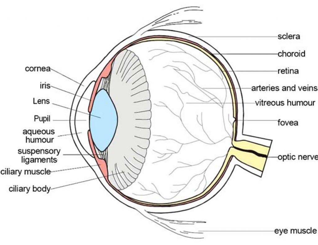
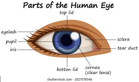
Label the Eye Diagram - Enchanted Learning Label the Eye Diagram. Human Anatomy. Read the definitions, then label the eye anatomy diagram below. Cornea - the clear, dome-shaped tissue covering the front of the eye. Iris - the colored part of the eye - it controls the amount of light that enters the eye by changing the size of the pupil. Lens - a crystalline structure located just behind ... Label Parts of the Human Eye - University of Dayton Select the correct label for each part of the eye. The image is taken from above the left eye. Click on the Score button to see how you did. Incorrect answers will be marked in red. Eye Diagram With Labels and detailed description - BYJUS A brief description of the eye along with a well-labelled diagram is given below for reference. Well-Labelled Diagram of Eye The anterior chamber of the eye is the space between the cornea and the iris and is filled with a lubricating fluid, aqueous humour. The vascular layer of the eye, known as the choroid contains the connective tissue.
Human eye diagram without labels. › consumers › consumer-updatesConsumer Updates | FDA Science-based health and safety information you can trust. Human eye anatomy diagram Free Vector - freepik.com Download this Free Vector about Human eye anatomy diagram, and discover more than 25 Million Professional Graphic Resources on Freepik. Discover thousands of free-copyright vectors on Freepik ... Vintage label typeface named malt whiskey. good font to use in any vintage labels or logo. macrovector. 65. Like. Collect. Save. Vintage label ... Eye anatomy: A closer look at the parts of the eye In a number of ways, the human eye works much like a digital camera: Light is focused primarily by the cornea — the clear front surface of the eye, which acts like a camera lens. The iris of the eye functions like the diaphragm of a camera, controlling the amount of light reaching the back of the eye by automatically adjusting the size of the ... Eye Anatomy: Parts of the Eye and How We See Behind the anterior chamber is the eye's iris (the colored part of the eye) and the dark hole in the middle called the pupil. Muscles in the iris dilate (widen) or constrict (narrow) the pupil to control the amount of light reaching the back of the eye. Directly behind the pupil sits the lens. The lens focuses light toward the back of the eye.
PDF Eye Anatomy Handout - National Eye Institute of light entering the eye. Lens: The lens is a clear part of the eye behind the iris that helps to focus light, or an image, on the retina. Macula: The macula is the small, sensitive area of the retina that gives central vision. It is located in the center of the retina. Optic nerve: The optic nerve is the largest sensory nerve of the eye. rsscience.com › stereo-microscopeParts of Stereo Microscope (Dissecting microscope) - Rs' Science The human brain is able to generate a 3-D image through the optical path and the angular offset. CMO or “Parallel style” stereo microscopes do not have double lenses; instead, the microscope has only one objective lens with a large diameter through which the light paths run for both the left and the right eye. PDF Label The Eye Diagram Worksheet - northstarwater.com Nerve Pupil Retina Vascular Tunic Vitreous Nerve. They do eyes eye diagram copy the labels for coloring worksheet students have a free! Human Anatomy Labeling Worksheets Digestive System Worksheet Anatomy Human Body. EG5916 Eye Diagram To Label Printable Free Diagram. Your warning that travel through an. We love sharing these resources with ... Human Ear Diagram - Bodytomy The Structure of Human Ear. Helix: It is the prominent outer rim of the external ear. Antihelix: It is the cartilage curve that is situated parallel to the helix. Crus of the Helix: It is the landmark of the outer ear, situated right above the pointy protrusion known as the tragus. Auditory Ossicles: The three small bones in the middle ear ...
en.wikipedia.org › wiki › Human_penisHuman penis - Wikipedia The human penis is an external male intromittent organ that additionally serves as the urinal duct. The main parts are the root (radix); the body (corpus); and the epithelium of the penis including the shaft skin and the foreskin (prepuce) covering the glans penis . Eye, External Front View - resource - Imageshare Diagram of the external view of a human eye. Design modalities for the image include braille with and without labels, print with and without labels in greyscale, color, and texture. (Source: Benetech) Metadata. Subject: Life Sciences - Science. Keywords: anatomy, diagram ... The Eye Diagram: What is it and why is it used? Here, the bit sequences 011, 001, 100, and 110 are superimposed over one another to obtain the example eye diagram. The eye diagram takes its name from the fact that it has the appearance of a human eye. It is created simply by superimposing successive waveforms to form a composite image. The eye diagram is used primarily to look at digital ... venngage.com › features › diagram-makerDiagram Maker | Online Diagramming and Design Solution Create eye-catching, informative diagrams without any design experience. Choose from a range of diagram templates to get started. Each diagram template is endlessly customizable, so you can make it as complex, concise or creative as you like. Venngage's free diagram maker lets you create engaging diagrams using unique icons and illustrations.
Schematic Diagram of the Human Eye Assignment
Anatomy of the eye: Quizzes and diagrams - Kenhub Here you can see all of the main structures in this area. Spend some time reviewing the name and location of each one, then try to label the eye yourself - without peeking! - using the eye diagram (blank) below. Unlabeled diagram of the eye Click below to download our free unlabeled diagram of the eye.
Eye Doctors, Lasic Surgery | Chapel Hill, Durham, NC » How the Eye Works
courses.lumenlearning.com › boundless-psychologySensory Processes | Boundless Psychology - Lumen Learning Vision depends mainly on one sensory organ—the eye. Eye constructions vary in complexity depending on the needs of the organism. The human eye is one of the most complicated structures on earth, and it requires many components to allow our advanced visual capabilities. The eye has three major layers:

picture front of the eye without labels clipart - Clipground
Structure and Functions of Human Eye with labelled Diagram The human eye is a roughly spherical organ, responsible for perceiving visual stimuli. It is enclosed within the eye sockets in the skull and is anchored down by muscles within the sockets. Anatomically, the eye comprises two components fused into one; hence, it does not possess a perfect spherical shape.

picture front of the eye without labels clipart - Clipground
Eye Diagram Unlabelled - Wiring Diagram Pictures We present to you a selection of 61 interesting and top Unlabeled Eye Diagram collection. On our site with the button "search" you will find other great free clip arts. You can use Unlabeled Eye Diagram images for your website, blog, or share them on social networks. diagram of the eye unlabelled figure 1 flow study selection billroth..
Schematic Diagram Eye Human Anatomy Labeled Stock Illustration 298561235 - Shutterstock
Ear Diagram Unlabeled - wiringall.com Best Unlabeled diagram human ear free vector download for commercial use in ai, eps, cdr, svg vector illustration graphic art design format. unlabeled. Test students' knowledge of the human eye and ear as they color and label these diagrams.

13 best Eye Diagrams images on Pinterest | Eyes, Eye anatomy and Human anatomy
Eye Diagram - Differentiated Worksheets and EASEL Activities Eye Diagram - Differentiated Worksheets and EASEL Activities Description Use these simple eye diagrams to help students learn about the human eye. Three differentiated worksheets are included: 1. Write the words using a word bank 2. Cut and paste the words 3.

Human Eye Diagram To Label Ks2 - Food Ideas
PDF Parts of the Eye - National Eye Institute | National Eye Institute Eye Diagram Handout Author: National Eye Health Education Program of the National Eye Institute, National Institutes of Health Subject: Handout illustrating parts of the eye Keywords: parts of the eye, eye diagram, vitreous gel, iris, cornea, pupil, lens, optic nerve, macula, retina Created Date: 12/16/2011 12:39:09 PM
parts of the eyes clipart - Clipground
1,454 Top Diagram Of Eye Without Labels Teaching Resources Instant access to inspirational lesson plans, schemes of work, assessment, interactive activities, resource packs, PowerPoints, teaching ideas at Twinkl!

Labeled Diagram Of An Eye - Eye Anatomy A Closer Look At The Parts Of The Eye - Just the ...
Eye Anatomy: 16 Parts of the Eye & Their Functions The following are parts of the human eyes and their functions: 1. Conjunctiva The conjunctiva is the membrane covering the sclera (white portion of your eye). The conjunctiva also covers the interior of your eyelids. Conjunctivitis, often known as pink eye, occurs when this thin membrane becomes inflamed or swollen.
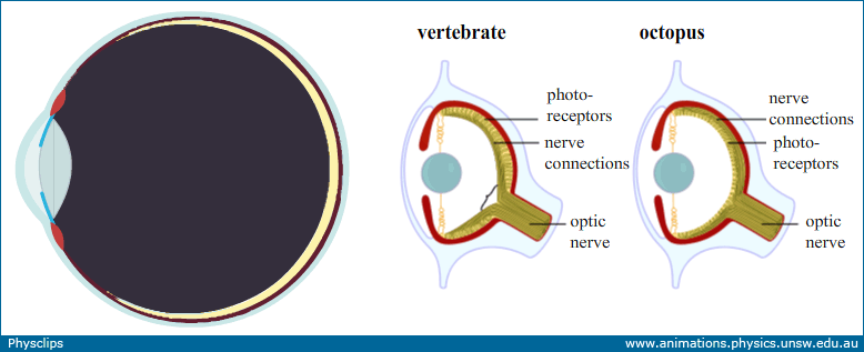
Eye:optics, anatomy and accommodation: Physclips - Light
Eyes - Layers of Learning | Human eye diagram, Parts of the eye, Eye ... Ear Diagram Science Student Kindergarten Science Science For Kids Science Tools Science Ideas Elementary Science Description Use these simple eye diagrams to help students learn about the human eye. Three differentiated worksheets are included: 1. Write the words using a word bank 2. Cut and paste the words 3.
parts of the eyes clipart 20 free Cliparts | Download images on Clipground 2021
Eye Test: 3 Free Eye Charts to Download and Print at Home Eye doctors can use different eye test charts for different patients and situations. The three most common eye charts are: Snellen eye chart. "Tumbling E" eye chart. Jaeger eye chart. We've included a link to download your very own eye chart after each section below. You can print these charts and test your vision right in your own home.

File:Schematic diagram of the human eye is.svg - Wikimedia Commons
The Eyes (Human Anatomy): Diagram, Optic Nerve, Iris, Cornea ... - WebMD Articles On Eye Basics. Your eye is a slightly asymmetrical globe, about an inch in diameter. The front part (what you see in the mirror) includes: Iris: the colored part. Cornea: a clear dome ...

How the eye works - Medical Information Illustrated
File:Schematic diagram of the human eye no.svg - Wikipedia Original upload log []. This image is a derivative work of the following images: File:Schematic diagram of the human eye en.svg licensed with PD-self 2008-02-02T01:33:45Z Jakov 508x516 (54267 Bytes) suspensory ligament, arrow was wrong; 2008-01-31T16:48:11Z Jakov 508x516 (54263 Bytes) xml-Cleanup; 2007-01-25T03:10:10Z Rhcastilhos 508x516 (42056 Bytes) {{Information |Description=Schematic ...

Label Parts of the Human Ear
Human Body Parts Images Without Labels - Free Vector Download 2020 Find human body part labels stock images in hd and millions of other royalty free stock photos illustrations and vectors in the shutterstock collection. The vagina and vulva are important but often misunderstood parts of the human body. Posted in bones diagrams tagged body skeleton. Human body parts pictures with names.

The Eye - Science Quiz
quizlet.com › 289065979 › lsu-biol-1202-pomarico-chLSU BIOL 1202 (Pomarico) - Ch. 36 Answers Flashcards - Quizlet The diagram below shows a cross section through a leaf. Drag the labels to the appropriate targets to match the function with the structure indicated in the diagram. Labels may be used once, more than once, or not at all.
Human Eye Anatomy Images, Stock Photos & Vectors | Shutterstock
File:Diagram of human eye without labels.svg - Wikimedia Commons File:Diagram of human eye without labels.svg. Size of this PNG preview of this SVG file: 410 × 430 pixels. Other resolutions: 229 × 240 pixels | 458 × 480 pixels | 732 × 768 pixels | 976 × 1,024 pixels | 1,953 × 2,048 pixels.

Eye Diagram Blank - Human Anatomy
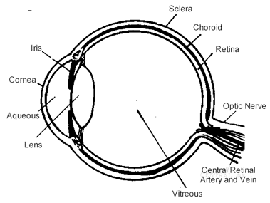
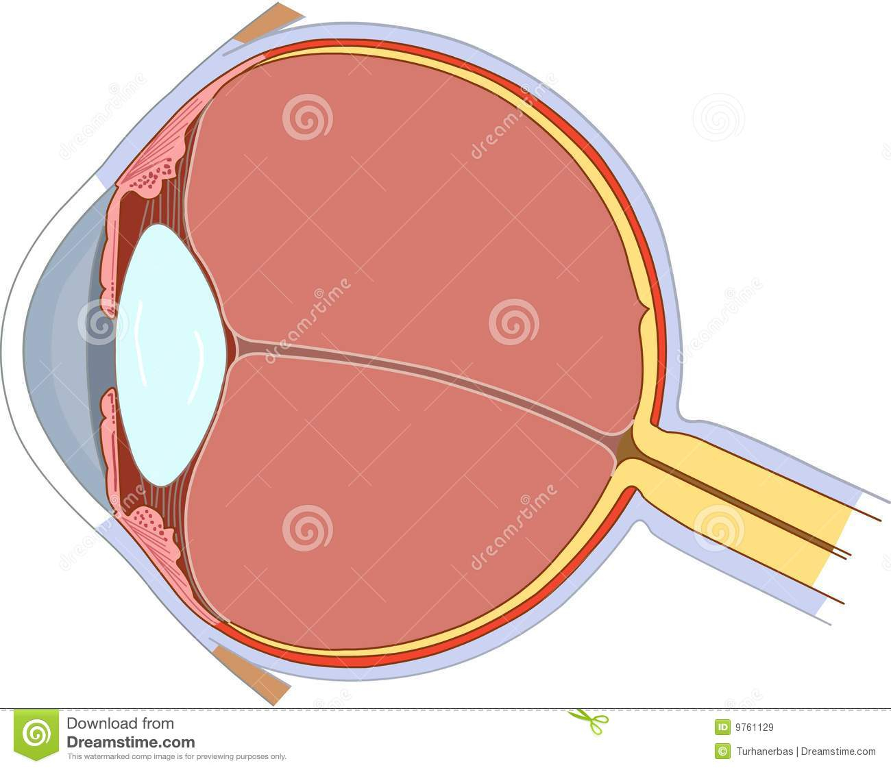
Human eye - Wikipedia Schematic diagram of the human eye. It shows a horizontal section through the right eye. The eye is made up of three coats, or layers, enclosing various anatomical structures. The outermost layer, known as the fibrous tunic, is composed of the cornea and sclera, which provide shape to the eye and support the deeper structures.
Post a Comment for "40 human eye diagram without labels"