39 light microscope with labels
Microscope Labeling - The Biology Corner 1) Start with scanning (the shortest objective) and only use the COARSE knob . Once it is focused… 2) Switch to low power (medium) and only use the COARSE knob . You may need to recenter your slide. Once it is focused.. 3) Switch to high power (long objective). Microscope, Microscope Parts, Labeled Diagram, and Functions Revolving Nosepiece or Turret: Turret is the part of the microscope that holds two or multiple objective lenses and helps to rotate objective lenses and also helps to easily change power. Objective Lenses: Three are 3 or 4 objective lenses on a microscope. The objective lenses almost always consist of 4x, 10x, 40x and 100x powers. The most common eyepiece lens is 10x and when it coupled with ...
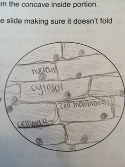
Animal Cell Diagram Under Light Microscope Labeled Tuesday, April 20th 2021. | Diagram. Animal Cell Diagram Under Light Microscope. To make observations and draw scale. This shows a generalized animal cell under a light microscope. We all keep in mind that the human physique is amazingly elaborate and one way I discovered to comprehend it is by way of the style of human anatomy diagrams.

Light microscope with labels
PDF Parts of the Light Microscope - Science Spot Supports the MICROSCOPE D. STAGE CLIPS HOLD the slide in place C. OBJECTIVE LENSES Magnification ranges from 10 X to 40 X F. LIGHT SOURCE Projects light UPWARDS through the diaphragm, the SPECIMEN, and the LENSES H. DIAPHRAGM Regulates the amount of LIGHT on the specimen E. STAGE Supports the SLIDE being viewed K. ARM Used to SUPPORT the Labeling the Parts of the Microscope Labeling the Parts of the Microscope This activity has been designed for use in homes and schools. Each microscope layout (both blank and the version with answers) are available as PDF downloads. You can view a more in-depth review of each part of the microscope here. Download the Label the Parts of the Microscope PDF printable version here. Animal Cell Under Light Microscope Labelled : Draw and label the ... Most cells are visible under a light microscope, but mitochondria and bacteria are barely visible. Record the microscope images using labelled diagrams or produce digital images. We say cells are microscopic because they can only be seen under a microscope. Note the whiplike flagellum that gives the cell a threadlike appearance.
Light microscope with labels. Compound Microscope: Definition, Diagram, Parts, Uses, Working ... - BYJUS The compound microscope is mainly used for studying the structural details of cell, tissue, or sections of organs. The parts of a compound microscope can be classified into two: Non-optical parts Optical parts Non-optical parts Base The base is also known as the foot which is either U or horseshoe-shaped. Microscope Drawing And Label - Painting Valley Label The Microscope... 270x350 14 0 Compound Light Micro... 630x380 8 1 Labeling The Parts O... 525x450 7 0 Compound Microscope ... 413x424 6 0 Microscope - Microsc... 236x262 4 1 Label Microscope Dia... 459x457 4 0 Microscope Parts Dia... 576x400 3 0 Section Cells View A... 512x346 2 1 Exe - Microscope Dra... 933x1163 2 0 Drawing - Microscope... Labeling the Parts of the Microscope | Microscope activity, Science ... Description Worksheet identifying the parts of the compound light microscope. Answer key: 1. Body tube 2. Revolving nosepiece 3. Low power objective 4. Medium power objective 5. High power objective 6. Stage clips 7. Diaphragm 8. Light source 9. Eyepiece 10. Arm 11. Stage 12. Coarse adjustment knob 13. Fine adjustment knob 14. Base Label the microscope — Science Learning Hub In this interactive, you can label the different parts of a microscope. Use this with the Microscope parts activity to help students identify and label the main parts of a microscope and then describe their functions. Drag and drop the text labels onto the microscope diagram.
Light Microscope- Definition, Principle, Types, Parts, Labeled Diagram ... A light microscope is a biology laboratory instrument or tool, that uses visible light to detect and magnify very small objects and enlarge them. They use lenses to focus light on the specimen, magnifying it thus producing an image. The specimen is normally placed close to the microscopic lens. Microscope Parts and Functions Microscope Parts and Functions With Labeled Diagram and Functions How does a Compound Microscope Work?. Before exploring microscope parts and functions, you should probably understand that the compound light microscope is more complicated than just a microscope with more than one lens.. First, the purpose of a microscope is to magnify a small object or to magnify the fine details of a larger ... Microscope Labeling Game - PurposeGames.com About this Quiz. This is an online quiz called Microscope Labeling Game. There is a printable worksheet available for download here so you can take the quiz with pen and paper. This quiz has tags. Click on the tags below to find other quizzes on the same subject. Science. Compound Microscope Parts, Functions, and Labeled Diagram Compound Microscope Definitions for Labels. Eyepiece (ocular lens) with or without Pointer: The part that is looked through at the top of the compound microscope. Eyepieces typically have a magnification between 5x & 30x. Monocular or Binocular Head: Structural support that holds & connects the eyepieces to the objective lenses.
Parts of Stereo Microscope (Dissecting microscope) - labeled diagram ... Labeled part diagram of a stereo microscope Major structural parts of a stereo microscope. There are three major structural parts of a stereo microscope. The viewing Head includes the upper part of the microscope, which houses the most critical optical components, including the eyepiece, objective lens, and light source of the microscope. Label the microscope — Science Learning Hub Jun 08, 2018 · All microscopes share features in common. In this interactive, you can label the different parts of a microscope. Use this with the Microscope parts activity to help students identify and label the main parts of a microscope and then describe their functions.. Drag and drop the text labels onto the microscope diagram. If you want to redo an answer, click on the … Compound Microscope Parts - Labeled Diagram and their Functions - Rs ... The eyepiece (or ocular lens) is the lens part at the top of a microscope that the viewer looks through. The standard eyepiece has a magnification of 10x. You may exchange with an optional eyepiece ranging from 5x - 30x. [In this figure] The structure inside an eyepiece. The current design of the eyepiece is no longer a single convex lens. ZEISS Axioscan 7 Microscope Slide Scanner Digitize your specimens with Axioscan 7 – the reliable, reproducible way to create high-quality virtual microscope slides. Axioscan 7 combines qualities that you would not expect to get in a slide scanner: high speed digitization and outstanding image quality plus an unrivaled variety of imaging modes are all available in a fully automated and easy to operate system.

Rough Endoplasmic Reticulum
Fluorescence - Wikipedia Fluorescence is the emission of light by a substance that has absorbed light or other electromagnetic radiation.It is a form of luminescence.In most cases, the emitted light has a longer wavelength, and therefore a lower photon energy, than the absorbed radiation. A perceptible example of fluorescence occurs when the absorbed radiation is in the ultraviolet …

Diagrams of Microscope | 101 Diagrams
Required practical - using a light microscope - BBC Bitesize Care must be taken when handling coverslips and microscope slides. Drawing the image Record the microscope images using labelled diagrams or produce digital images. When first examining cells or...

Quia - 9AP Chapter 12 - The Cell Cycle (Detailed)
Microscope Labeling - The Biology Corner Students label the parts of the microscope in this photo of a basic laboratory light microscope. Can be used for practice or as a quiz. Name_____ Microscope Labeling . Microscope Use: 15. When focusing a specimen, you should always start with the _____ objective.

Histology Drawings: January 2014
Solved Label the image of a compound light microscope using - Chegg Experts are tested by Chegg as specialists in their subject area. We review their content and use your feedback to keep the quality high. Transcribed image text: Label the image of a compound light microscope using the terms provided.
Using the Compound Microscope in Class - Microscopy
An Introduction to the Light Microscope, Light Microscopy Techniques ... a) standard upright microscope indicating (1) e yepiece (ocular lens), (2) objective turret, revolver, or revolving nose piece (to hold multiple objective lenses), (3) objective lenses, focus knobs (to move the stage) (4) coarse adjustment, (5) fine adjustment, (6) stage (to hold the specimen), (7) light source (a light or a mirror), (8) …

Microscope World Blog: Bacteria under the Microscope
Light Microscope: Functions, Parts and How to Use It The function of the light microscope is based on its ability to focus a beam of light through a very small and transparent specimen, to produce an image. The image is then passed through one or two lenses for magnification to view. The transparency of the specimen allows for easy and fast light penetration. Specimens can vary from bacteria to ...

Microscope World Blog: Cheek Cells under Phase Contrast Microscope
Simple Microscope - Diagram (Parts labelled), Principle, Formula and Uses A simple microscope consists of Optical parts Mechanical parts Labeled Diagram of simple microscope parts Optical parts The optical parts of a simple microscope include Lens Mirror Eyepiece Lens A simple microscope uses biconvex lens to magnify the image of a specimen under focus.

Prokaryote Under Microscope
Parts of a microscope with functions and labeled diagram Microscopic illuminator - This is the microscopes light source, located at the base. It is used instead of a mirror. It captures light from an external source of a low voltage of about 100v. Condenser - These are lenses that are used to collect and focus light from the illuminator into the specimen.
Post a Comment for "39 light microscope with labels"